

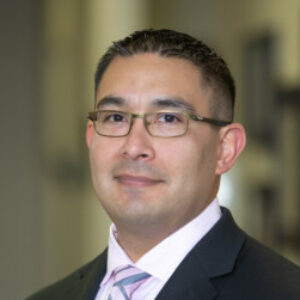
53 year old male with right lower extremity pain. We performed a right L5/S1 transforaminal discectomy and decompression. Patient was found to have a calcified disc and we were able to successfully decompress the L5 nerve and patient had alleviation of right lower extremity pain.
Transcript
3,674 words · 20 min
Transcript
3,674 words · 20 min
I'm Doctor Domingo Molina. I'm an endoscopic Spine surgeon. This is a right L5-S1 transaminal discectomy. We will be discussing indications and technique. We'll save targeting and localization for another video.
This is a 53 year old male presented to the clinic with complaints of right Hip Pain. The patient had exhausted conservative care since 2013, had undergone injections, and was still having Pain. He wanted a minimally invasive Surgery and wanted to get back to an active lifestyle pretty quickly. So, um, we indicated a right L5-S1 transframinal discectomy. If we look here at the.
X-rays, uh, you notice the crest is fairly low.
So, it's a perfect uh.
Perfect anatomy for a right L5 transaminal discectomy. I typically check the crest on the AP and laterals. If the crest is below the pedicle of L4, then it's usually amenable to a transraminal discectomy.
If we look here at the T2 sagittal MRI, you notice a little bit of.
Disc bulge into the framing of the L5-S1 disc space. Uh, you can notice some ligamentum flavum coming off the SAP as well, so, so much stnotic uh foramen.
The T2 axial is typically where the money is at. The L5-S1 frame and on the right side here, a little bit of a hypertrophic facet, uh, you notice the transfframinal uh disc herniation, a little bit of a far lateral nature to it, a little bit of a high intensity zone into the uh paracentral region of the uh L5-S1 disc.
So I'll kind of go over my initial starting point for endoscopic, uh, transramal discectomy. Uh, so we used a transSAP Approach in this procedure. Uh, that means docking the GM sheeting onto the kind of most cranial aspect of the super articular process of S1 and then advancing remers down into it.
And so what that does is it uh removes the uh most cranial aspect of the SAP and enlarges the foramen, uh, so we can do most of our work. So what we're looking at here, uh, the cranial aspect of the, um, camera is the
Uh, is basically the framing that I created, uh, with the reamers right now, I'm removing a lot of the fat tissue, epidural fat.
As well as um just some of the uh ligamentum flavm from the uh facet joint.
So where we move to now is that you can see the superior articular process in the uh upper left-hand corner. I've got the RF probe. I'm checking my pedicle um of the uh S1 vertebral body, making sure that I'm in the correct location. I'm using a fluoroscopy and AP plane for this. And I'm trying to remove some of this, uh, ligament attachment from the, uh, facet joint. And that's really what you see on the MRI that I showed initially, and that's where you can get some.
Uh, foraminal compression when people stand, um, they most likely complain of leg Pain, uh, when standing, and so the foraminal area of the.
Uh, the femoral area gets smaller and causes compression radiculopathy. And so, uh, as you can see here, this is a perfect view. Uh, we removed the suparticular process tip. Um, there's still some of the supra articular process that we're going to address. Um, and so what I'm doing right now is just removing some of this fat out of my, uh,
Out of my working view, uh, you can kind of see once I remove the instruments, you can kind of see the canal pulsations come through with the epidural fat on the right corner. And, uh, so what I'm gonna do is I'm gonna take the diamond burn and I'm gonna remove some of this super articular process here in my way and, uh, further enlarge the foramina.
And so what I do is I kinda uh like to start kind of inside out. And so what I'll do is I'll advance it kinda where you see here, and then I'll kinda pull it back. And so, this is safer rather than plunging into the canal. Uh, it gives you a little bit more control cause I can kinda feel that lip right there.
And then pull it back.
And like I said, it uh keeps the traversing nerve roots safe. So, um, you can see some of that ligament attachment on the Other side.
And so again, uh, just kind of removing some of the super articular process here. This burr runs at 10,000 RPM, so, uh, it doesn't take a lot to remove a lot of this bone. Uh, you would think that you'd have to do a lot of bony work. Um, but again, you're working kind of in a small area and, um, you know, in Spine Surgery, millimeters count, and so, um,
You know, sometimes these patients don't need a, a ton of extra room, um, but you do need to kind of adequately, uh, decompress, um, all the structures here. So, um,
So again, still taking the uh the burr here, I'm kind of trying to remove some of the ligandum flavor attachment with the burr as well.
So we must be finished with our burr work here.
As you can see, there may be a nerve on the Other side of that, um, you know, it may also still be a little bit of the ligamentum flavum as the lateral recess comes down. So you kinda wanna be careful plunging into that area there. I usually use an RF probe to kind of probe the areas, um, to make sure, again, I'm.
Uh, kind of where I need to be, so I always use an RFPR for localization. It's almost done with the burr work here.
Again, you can kind of see I've taken off uh.
A fair amount of the SAP, um, but not so much that it destabilizes anything. Uh, you know, it's, it's a little bit of just experience on, on how you determine how much you need. I kind of, um, try to access a little bit of the disc space as well through that and make sure that the, uh, exiting nerve root is decompressed. So here, I'll take the kerosene, try to remove some of the ligamentum, uh, attachment, uh, from the SAP again, and try to just remove any sharp edges.
Again, uh, a lot of these, uh, foraminal stenosis cases, the tip of the SAP as well as the ligamental inflamm is what impinges on the nerve root, as well as the disc bulging anteriorly, so.
Uh, in this case, uh, you know, we'll, we'll talk about the disc herniation, uh, towards the end of the case. Um, but right now, my goal is to, uh, really decompress the foramina, really get a good idea of where I'm at, uh, maybe visualize the nerve.
And um
You know, uh, again, uh, visualize the disc space, and once I do that, then I can kind of see what else I need to decompress. Uh, so, again, here, I almost got the ligament attachment off, and this is gonna kind of really open up our window here for everything. So, uh, these kerasins, unfortunately, are not super robust, and so you wanna kinda do most of your bony work with the burr if you can.
Uh, the kerisons are not the normal sized kerisons, um, unless you're using.
Uh, the older traditional join max probe, or it's gonna be a working tube. So, um, again, here's my, uh, pituitary, I'm removing a lot of this. This ligamentum was really sticky, uh, so it was, uh, really hard to remove a lot of this.
Uh, another question may be, well, how do you know what you're grabbing? Well, as long as they stay within my working tube, I know where the nerve is. I know that the exiting nerve is to the right of the working tube, um, protected under the
Uh, kind of near the pedicle. Um, I know that, uh, I'm right under the SAP and so anything within my working tube I can grab. As far as depth, I know that I'm not in the canal, so, um, again, you kinda wanna not try to stray out too far from the working tube. There are certain bites that I do use that for, uh, but again, um, I kinda really know where I'm going. Um.
And so here in the bottom corner, you can kind of see this is where the case gets interesting. Um, this patient ended up having a Calcified disc herniation. Um, I don't typically get uh CT scans on, uh, lumbar cases. Um,
It's probably a good rule of thumb if the patient has had radicular symptoms for even more than a year or close to a year to probably get CT scans. I'm pretty comfortable that once I get in there, it's not really gonna change my treatment. I'm going to either shave down the calcified disc and open up the frame, and a lot of these patients, I don't indicate a fusion on, and I'm doing this, uh, endoscopic case just solely to, uh, you know, for.
Uh, conservative purposes, so I'm gonna do some sort of decompression again, whether it's a soft disc or a calcified disc. Um, some people may agree with that, some people not, but I think that's the nice thing about the Join max is, uh, the ability to do that, so.
So we're kind of fast forward for uh time purposes here, so.
Um, we've already removed the tip of the super articular process. I'm using the RF probe right now to try to localize, uh, actually where the disc space is. And so, in this case, uh, you know, I took an AP radiograph, I took a lateral radiograph. I'm trying to check where the pedicle is. Um, I know that I'm over the disc space, um, and so I'm really just trying to be careful and.
Kind of figure out where I'm at. You can kind of see some of the traversing kind of epidural veins as well as um the traversing nerve root is right uh behind that, so.
Um, sometimes a little bit hard to differentiate some of these structures, um.
You know, you can kind of uh use the bipolar setting rather than a vape setting and kind of touch it to any of the structures to see if uh you get any EMG activity. I do monitor all these cases with EMG and so, that's always uh.
Good to do, um.
So here, I'm kinda looking at the joint, um, kind of seeing it move, making sure I can, uh, again, visualize where I'm at in my head. Um, there's still a little bit of ligamentum attachment. Um, I'm not too apt to get this. I'm trying to kind of, uh, remove it with, uh, the, the vape setting on RF probe. Um, again,
Um, you know, traditionally, you should kind of see the traversing nerve in the canal. This patient did not have lateral recess stenosis symptoms. Uh, so, again, I'm really trying to clean up the foraminal window, um, you know, rather than the lateral recess. Um, sometimes using your trajectory, uh, is good and bad. If your trajectory is, uh, too high, if you come in kind of too, uh, cranial to coddle.
Then you won't be able to ever get into the lateral recess. I've had this situation before. And so here, um, I'm kind of putting in a lot of these slides, uh, to, to show you, uh, really, truly how I do this and try not to
To leave anything out, uh, for people that are just getting into Endoscopic spine surgery. And so, you know, you see a lot of bleeding here. What I do is I kind of push the camera down really close to the
To the Pathology and um that'll kinda help me try to visualize where the bleeding is coming from. I use Epi um in my bags of saline. Um, I actually just started doing that. It's been a real difference. What I'll do is I'll take the bipolar probe, kind of probe around here. Um, what I had done is I had stuck our probe, uh, kind of to eventually and, um,
If you know, from open procedures, that's not good because that's kind of where you get into those really.
Brisk bleeders and so.
Um, so eventually I was able to control it, um, stick in our probe.
Uh, down in there, um.
And you, you kind of wanna really hold it over over what's bleeding too. I know that seems a little bit intuitive, but uh.
you know, sometimes it's not when you're actually doing this, um, in a person. So, sometimes even some of these bleeders from this epidural fat can, um, can really get you. So, um, so it's really important to get those two. Again, um, you know, I don't know if you saw a hint of the exiting L5 nerve root there on the side. That's the nice thing about the JoyMax cameras, you can really see it. And so,
If you look here, um, I finally was able to kind of figure out the disc space, um, it's calcified. I'm kind of removing some of this fat, but if you saw I could press with the RF probe and, um, the annulus, uh, it was kind of moving a little bit, it's a little bit spongy still, but definitely a calcified, uh, component to it over it. So, um, so what I'm gonna do is, um, you know, here at this point, I was kind of thinking, well, do I just leave the decompression as it is, or do I
Go ahead and take the Diamondbuur and.
Uh, decompress it.
So, at this point I'm using a diamond bird to decompress the calcified disc. I'm basically.
Just, uh, removing enough that way I can get into the disc space a little bit, remove some of that far lateral kind of transaminal component that we had seen. Uh, in a minute, we're gonna take a look at the exiting nerve root. Uh, but I do know that the exiting nerve root is free. Um, and so, how much do I remove here? Um, honestly, it's just kind of a guess. Um, I'm using fluoroscopy in the lateral plane to check my depth. Um, I'm gonna see here, I kinda start moving the disc space a little bit.
Uh, checking for.
Uh, the annual is, uh, because I know that the whole thing is not totally calcified.
And so, um, I can see a little bit of some, you know, fibers of the annulus there, um.
And so I know that the SAP was really the main culprit as well as the hypertrophic ligamentum flavum attachment off of the SAP, um, and so, um, I'm not gonna spend too much time doing this, and I'm already towards the end of the case here, um, but just wanna again, make sure that I do have a thorough decompression. That way, when the patient stands, he doesn't have any radicular symptoms from the compression.
And the decrease of uh the foraminal area.
Um, so I'm getting a little bit far out here. How do I know that? Um, you know, I, I just know that, um, where my working tube is. I don't wanna stray out too far laterally, um.
You know, cause you always kinda wanna keep, uh, keep the work to where you can recognize anatomical structures, um, and know where you're at. Um, so you can see here, we're getting more of the annulus fibers here. Um, the disc is pretty chalky. Um, and so that's, that's what I'm removing with the, with the diamond burr here. I am in a disc space here. I'm taking fluoroscopic shots at the, at the same time, uh, that you can't see, but I'm checking my depth, um, just to kind of one annotate that I'm in the disc space.
Um, and then 2, making sure that I do, uh, remove enough of the, of the disc space, uh.
Just to have a good thorough decompression. So this time, I'm using my pituitary to remove some of the calcified discs and some of just the chalky annulus fibers. I'm not gonna spend too much time doing this, um, because it's again, that's not really the, the main culprit of my, uh, compression, um.
Especially since it's calcified, I know that, uh, if the patient continues to have radicular symptoms, you know, I'll, I'll talk to him about uh possible effusion later. But again, I've had several of these cases where usually decompressing a calcified disc and uh doing some of the, uh, bony work, uh, really alleviates a lot of their symptoms and, uh, you know, we can get them out as far as possible, uh, before having a fusion. But I do counsel them postoperatively and say, hey, the, the disc is calcified.
The level's very degenerative, um, you know, we did take care of your symptoms now, but, um, you know, there may be a, a fusion, um, in your future.
Um.
I had a guy with a, with a L4 um quad palsy, uh, that after my decompression, I wasn't so sure that I'd gotten everything just because of the degenerative nature, but, um, again, uh, he ended up getting full quad function back. Um, he was 86, I believe, and so.
Um, again, um, I control some of this bleeding again, uh, with the RF probe, um.
You know, you, you wanna make sure that you control some of the bleeding just to make sure you don't have any epidural hematomas. Following that, I have had a couple of cases, um, where, you know, the water pressure will, will kind of stave off some of the epidural bleeding, and then you take the tube out, and then later on, they have continued radicular symptoms or, uh, they have an epidural hematoma because, uh, there wasn't as much bleeding as you thought there was, so.
Um, so this is, uh, kind of where we, uh, kinda check our decompression. Again, um, I didn't get really into the canal here. Um, you know, I still haven't in my cases really, unless they have a lot of recess symptoms, um, really spent a lot of time decompressing that area, uh, mostly because I don't like to take blind bites into the canal to try to, there's usually a lot of epidural fat, um, but I can see the canal pulsations there.
Um, you can see here that the SAP is removed, um, or at least the tip of it, and this is a great view of the L5 exiting nerve. And so I couldn't be happier with, um, getting this decompression. I know that his radicular symptoms are most likely gonna be gone because again, he's got a ton of room in here now for the exiting L5 nerve root. There's a little bit of some disc right there, and so the great thing about the working tube is that I can kind of, um,
Push the nerve out of the way. I didn't wanna go the Other way, so I'm coming back this way cause I don't wanna squish the uh L5 nerve or compress it. I guess squish is a medical term. But, um, you know, the L-five nerve can sometimes be very tenuous, and so, I don't wanna push on it too much. Um, again, this is just kind of an experience, uh, feeling, uh, on how much to, to retract the nerve root. Again, I'll use the pituitary to remove that little bit of some, uh, disc there again. Um.
And so, you know, again, we're really not gonna spend too much time going to that corner because even though you can kind of see it, you don't wanna get too far into there cause the nerve does take a pretty oblique angle at the L5 S1 disc space. And so, um, that's really been evident, uh, when I've done these cases really to learn the anatomy of the exiting nerve root and, um, and so,
Uh, you know, it's been really great to actually see, well, how does the L5 nerve root travel. So, again, great view here. Everything's decompressed. The SAP is removed. I removed some of the calcified disc. Um, the L5 nerve looks great. It's free and, uh, clear. I know that when this patient gets up, he's not gonna have any ridiculous symptoms. This foraminal area is going to collapse a little bit when the patient stands up. And so again, that's where some of that hypertrophic ligamentum flavm and that facet.
Um, or the SAP can really compress on a nerve root, and so you can imagine that if the full SAP was there, um, as it was initially, you can understand now how, um, it can be compressed and cause radicular symptoms.
So that was the end of our case. Thank you for watching and listening. I'll try to post some more of these videos and kind of new techniques, uh, for people that are getting into endoscopic Surgery. Uh, the patient did have a complete alleviation of his uh radicular Hip symptoms and, uh, Pain down the right lower extremity. Following that, uh, less than a 1 centimeter incision, sent patient home, uh, approximately an hour and a half after Surgery. So, my prosop Protocols are typically no extreme heavy bending, twisting, or lifting.
Uh, for a couple of weeks and the patient can, uh, especially if there's no disc component to it, they can get back to activity a little bit quicker. Um, if anybody has any questions, just, uh, send me a message and I'd be happy to, um, either give advice or explain things further. Thank you.
Automatically transcribed and not checked by a person — it may contain errors, particularly in names, drugs and dosages. The recording is the source.
Check yourself
5 questions5 questions on what was actually said in this recording, and we remember how you did. Sign in to take it — an account is free, and nothing else on doc.social asks for one.
AI-generated from this recording’s transcript, not written by the speaker. Tell us if a question misreads what was said.
Related lectures
 Lecture
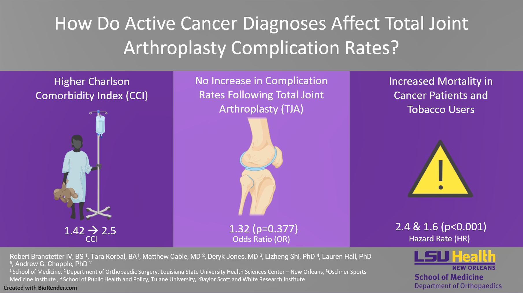
How Do Active Cancer Diagnoses Affect Total Joint Arthroplasty Complication Rates?
There is currently a paucity of data analyzing total joint arthroplasty (TJA) disparities among patients with active cancer diagnoses.
Lecture
Targeted Nutrition in Orthopedic Surgery - A Missed Opportunity to Enhance Outcomes
Surgery triggers a series of inflammatory, immune, and metabolic responses. The stress response leads to an increase in protein breakdown that outpaces protein synthesis. The body's natural store of essential amino acids exit skeletal muscl…
 Lecture
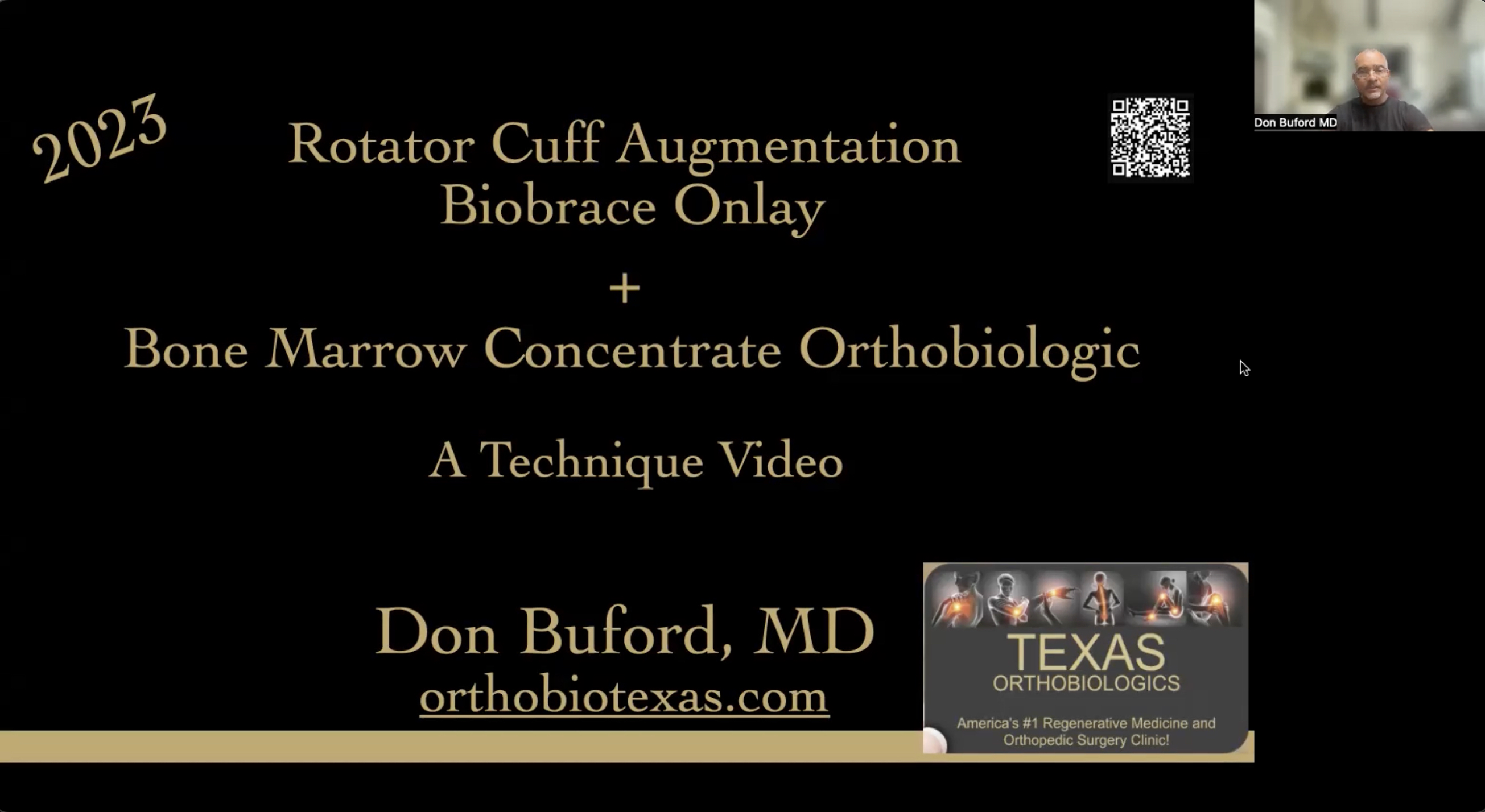
Rotator Cuff Repair with Biobrace augmentation and Bone marrow concentrate
Take a look at how to increase your success rate for healing a torn rotator cuff!